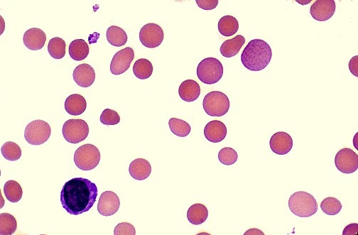
Acanthocytosis

Retinitis pigmentosa showing bone spicule pigmentation associated with acanthocytosis and abetalipoproteinemia

Acanthocytes, also known as spur cells, are abnormal red blood cells characterized by irregularly spaced, variably sized membrane projections, reflecting a true structural alteration of the erythrocyte membrane rather than a reversible shape change. The development of acanthocytosis results from disruption of the lipid composition and reduced fluidity of the red cell membrane, most commonly due to an increased cholesterol-to-phospholipid […]

Read more

Stomatocytosis

Peripheral blood smear image showing red blood cells with a central slit-like pallor, characteristic of stomatocytosis.

Stomatocytosis is a rare red blood cell (RBC) membrane disorder characterised by the presence of stomatocytes—red cells with a slit-like or mouth-shaped central pallor—on the peripheral blood smear. Hereditary stomatocytosis and acquired forms are associated with altered membrane permeability, hemolysis, and variable degrees of anemia. Recognition of stomatocytosis is important in hematology because it may indicate an underlying red cell […]

Read more

Elliptocytosis

Peripheral blood smear showing elliptical red cells (elliptocytes) characteristic of hereditary elliptocytosis.

Understanding Hereditary Elliptocytosis (HE): A Defect of the Red Cell Membrane Hereditary Elliptocytosis (HE), or ovalocytosis, is defined as a genetic disorder of the red cell membrane that affects the shape of red blood cells. Characterized by the presence of a majority of elliptical-shaped cells on the peripheral smear, HE results from specific mutations in cytoskeletal genes, notably spectrin (SPTA1, […]

Read more

Spherocytosis

Spherocytes in warm autoimmune hemolytic anemia showing small dense red blood cells with loss of central pallor

Hereditary spherocytosis (HS) is the most common inherited red blood cell membrane disorder, typically transmitted in an autosomal dominant pattern. It is characterized by structurally abnormal red blood cells that become spherical (spherocytes), rigid, and osmotically fragile. On peripheral blood smear, these cells appear smaller, dense, and deeply stained with loss of central pallor. While spherocytes may be seen to […]

Read more

Hemolytic Anemias

Close-up of eyes showing pronounced scleral icterus due to hyperbilirubinemia in hemolytic anemia

Introduction: Red blood cells (RBCs) are produced in the bone marrow and normally survive in the circulation for approximately 120 days before being removed by the reticuloendothelial system. Hemolysis refers to the premature destruction of red blood cells, resulting in a shortened RBC lifespan of less than 120 days. In response, the bone marrow increases erythropoiesis to compensate for the […]

Read more

Pure Red Cell Aplasia

Bone marrow smear in Pure Red Cell Aplasia showing severe erythroid hypoplasia with preserved myeloid cells and a single late erythroblast indicated by a yellow arrow.

Introduction: Pure Red Cell Aplasia (PRCA) is a rare hematologic disorder characterized by a selective failure of erythropoiesis, resulting in profound anemia with markedly reduced or absent reticulocytes. Although erythroblasts are virtually absent in the bone marrow, granulopoiesis and thrombopoiesis remain intact, allowing white blood cell and platelet counts to stay within normal limits. The anemia is typically normocytic but […]

Read more

Aplastic Anemia

Bone marrow biopsy showing marked hypocellularity with extensive fatty replacement, characteristic of aplastic anemia on H&E staining.

Aplastic anemia is a rare and serious bone marrow failure disorder in which hematopoietic stem cells become damaged, leading to markedly reduced blood cell production. As a result, patients develop pancytopenia, characterized by anemia (low red blood cells), leukopenia (low white blood cells), and thrombocytopenia (low platelets). The term aplastic refers to the marrow’s inability to generate mature, functional blood […]

Read more

Megaloblastic Anemias

Atrophic glossitis showing a smooth erythematous tongue with loss of papillae in vitamin B12 or folate deficiency

Introduction: Megaloblastic anemias represent a spectrum of disorders caused by impaired DNA synthesis affecting all rapidly dividing hematopoietic cells, not only the erythroid lineage. When DNA replication is disrupted, the cell cycle cannot progress normally from the G2 phase to mitosis, resulting in continued cytoplasmic growth without division. This nuclear–cytoplasmic asynchrony produces the hallmark morphological features of macrocytosis and megaloblastosis. […]

Read more

Iron Deficiency Anemia

Clinical sign of pallor and paleness in a female patient with Iron Deficiency Anemia

  Iron deficiency anemia (IDA) is the most common type of anemia, and it occurs when your body doesn’t have enough iron. Your body needs iron to make hemoglobin. When there isn’t enough iron in your bloodstream, the rest of your body can’t get the amount of oxygen it needs. Iron is a mineral. Most of the iron in the […]

Read more

Anemias-General Approach

Anemia symptoms showing fatigue weakness and difficulty concentrating

Anemia is a common hematological condition characterized by a reduction in circulating red blood cells or hemoglobin (Hb), resulting in impaired oxygen delivery to body tissues. It is not a diagnosis in itself but rather a clinical manifestation of an underlying disorder; therefore, even mild or asymptomatic anemia warrants proper evaluation to identify and treat the primary cause. When anemia […]

Read more
1 7 8 9 10