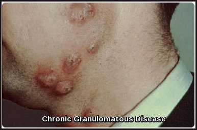
Langerhan’s Cell Histiocytosis

Extensive erythematous skin lesions on the chest in a patient with Langerhans Cell Histiocytosis

Langerhans Cell Histiocytosis (LCH), historically referred to as Histiocytosis X, represents a spectrum of clonal dendritic cell disorders ranging from the typically localized and indolent unifocal eosinophilic granuloma to the more aggressive multifocal form and the fulminant disseminated variant previously known as Letterer–Siwe disease, which carries a high risk of morbidity and mortality. This heterogeneous group of conditions is characterized […]

Read more

Gaucher’s Disease

Bone marrow smear showing characteristic Gaucher cells with wrinkled tissue-paper cytoplasm in Gaucher’s disease.

Gaucher’s disease is a rare inherited lysosomal storage disorder caused by deficient activity of the enzyme glucocerebrosidase. This autosomal recessive condition results from pathogenic variants in the GBA gene, leading to the accumulation of glucocerebroside within macrophages. These lipid-laden “Gaucher cells” progressively infiltrate the liver, spleen, bone marrow, and—in some subtypes—the central nervous system, producing a wide spectrum of clinical […]

Read more

Sea Blue Histiocytes

Microscopic image showing a sea blue histiocyte in bone marrow with dense blue cytoplasmic granules typical of sea blue histiocytes.

Sea Blue Histiocytes are enlarged, lipid-laden macrophages found in the bone marrow that characteristically exhibit a deep sea-blue cytoplasmic staining on Romanowsky preparations. These distinctive cells arise when histiocytes accumulate phospholipids or other lipids that resist normal degradation pathways, giving them their granular blue appearance under light microscopy. Although Sea Blue Histiocyte Syndrome is a rare autosomal dominant condition associated […]

Read more

Chediak-Higashi Syndrome

Child with Chediak-Higashi Syndrome showing partial oculocutaneous albinism and pigment dilution

Chediak-Higashi Syndrome (CHS) is a very rare autosomal recessive primary immunodeficiency disorder characterized by a complex mutation in a lysosomal trafficking regulator protein. This mutation severely impairs the immune system’s function (phagocytosis and bacterial lysis), leading to devastating consequences for the patient, including recurrent bacterial infections (especially respiratory), partial albinism, an increased bleeding tendency, and often progressive neurological disease. While […]

Read more

Chronic Granulomatous Disease

Cutaneous granulomatous lesions on the face and neck in a patient with Chronic Granulomatous Disease (CGD)

Chronic Granulomatous Disease (CGD) is a rare inherited primary immunodeficiency in which phagocytes fail to generate reactive oxygen species (ROS) due to defects in the NADPH oxidase enzyme complex. As a result, affected individuals are highly susceptible to recurrent, severe bacterial and fungal infections and may develop granulomatous inflammation in various organs. The study of CGD has been pivotal in […]

Read more

Lupus Erythematosus (LE) Cells

LE cell in systemic lupus erythematosus showing neutrophil engulfing denatured nuclear material

Lupus Erythematosus (LE) cells are characteristic neutrophils that have phagocytosed the denatured nuclear material of damaged lymphocytes, typically coated with autoantibodies against nucleoproteins. First identified by Dr. Robert H. Heitzman in 1948, LE cells once served as a key diagnostic marker for systemic lupus erythematosus (SLE), reflecting the autoimmune pathophysiology of the disease. Although largely replaced today by more sensitive […]

Read more

Pelger-Huët Anomaly

Blood smear showing bilobed neutrophils characteristic of Pelger-Huët anomaly

Pelger-Huët Anomaly (PHA) is a rare, typically benign hereditary disorder of neutrophil nuclear segmentation, characterised by bilobed or peanut-shaped nuclei instead of the normal multilobed appearance. This distinctive morphology affects neutrophils and other granulocytes and is often discovered incidentally on a peripheral blood smear. Although most individuals with true (inherited) PHA remain asymptomatic with normal immune function, similar morphological changes—termed […]

Read more

Leishmaniasis

Cutaneous leishmaniasis ulcer on the hand showing inflamed borders and central tissue loss

Leishmaniasis is an infectious disease caused by intracellular protozoan parasites of the Leishmania genus, transmitted to humans through the bite of infected female sand flies. The condition occurs across several endemic regions worldwide and can present in cutaneous, mucocutaneous, or visceral forms. More than 20 Leishmania species are capable of causing human disease; although they appear morphologically similar, they are […]

Read more

Lymphatic Filariasis

Three patients with advanced lower-limb lymphedema caused by chronic filariasis.

Lymphatic filariasis is a mosquito-borne parasitic infection classified as a neglected tropical disease (NTD) and caused by microscopic thread-like filarial worms that reside in the human lymphatic system. This system is essential for maintaining fluid homeostasis and immune defence, and chronic infection can lead to significant lymphatic dysfunction. Transmission occurs when infected mosquitoes introduce larval parasites (microfilariae) into the bloodstream, […]

Read more

Trypanosomiasis

Trypanosomiasis

Trypanosomiasis, also known as sleeping sickness or Chagas disease, is a vector-borne parasitic infection caused by protozoa of the genus Trypanosoma. It affects both humans and animals and is transmitted primarily through the bite of infected tsetse flies (Glossina species) in Africa or by triatomine (kissing) bugs in the Americas. The African form is caused by Trypanosoma brucei gambiense (West […]

Read more
1 5 6 7 8 9 10