Lupus Erythematosus (LE) Cells
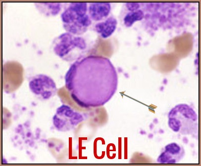
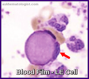
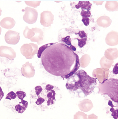
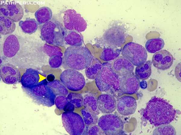
Lupus Erythematosus (LE) Cells are neutrophils that have engulfed lymphocyte nuclei coated with and denatured by antibody to nucleoprotein. These cells were first described by Dr. Robert H. Heitzman in 1948, and they play a significant role in diagnosing systemic lupus erythematosus (SLE).
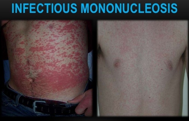
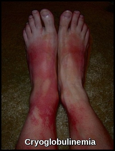
The Lupus Erythematosus (LE) cell was so termed because of its exclusive presence in the bone marrow of 25 patients with confirmed or suspected SLE at the Mayo Clinic. The first three cases were children, the first a 9‐yr‐old with, very unusually, hypergammaglobulinaemia, plasmacytosis and Bence‐Jones proteinuria; the second, also 9‐yr‐old, with unexplained anaemia and purpura, and the third a boy with cryoglobulinaemia and purpura.
A more conclusive link with SLE came after the LE cell phenomenon was observed in the marrow of a patient with classical SLE in 1948 by Hargraves et al.
Lupus Erythematosus (LE) Cells are not usually found in peripheral blood, although Sundberg and Lick, also at the Mayo Clinic, observed in 1949 that the LE cell phenomenon could form in the buffy coat of peripheral blood after a period of incubation at room temperature while rotating with glass beads.
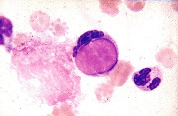
A blood smear from a patient with systemic lupus erythematosus shows a classical LE cell in which a viable neutrophil has ingested nuclear material. Note the nuclear debris and an adjacent normal neutrophil and the nuclear debris. Courtesy of the American College of Rheumatology.
Lupus Erythematosus (LE) Cells have since been found in synovial fluid, cerebrospinal fluid and pericardial and pleural effusions from patients with SLE.
The LE cell reaction is positive in 50%-75% of individuals with acute disseminated systemic Lupus Erythematosus (SLE). Positive reactions are also seen in rheumatoid arthritis, chronic hepatitis, scleroderma, dermatomyositis, polyarteritis nodosa, acquired hemolytic anemia, and Hodgkin’s disease. It may also be positive in persons taking phenylbutazone and hydralazine.
Lupus erythematosus (LE) cell testing was once performed to diagnose systemic lupus erythematous but has been replaced for this purpose by antinuclear antibody testing.
Negative findings on Lupus Erythematosus (LE) cell testing exclude a diagnosis of systemic lupus erythematosus (SLE).
The presence of LE cells indicates lupus.
A smear is considered positive when ten or more characteristic LE cells are seen during a 15-minute search associated with the presence of extracellular, amorphous, nuclear masses.
The discovery of the LE cell phenomenon remains a fundamental observation in the pathogenesis of SLE. Along with the VDRL test, the LE cell preparation remains in the American College of Rheumatology’s criteria for the classification of SLE.
It’s important to note that while the presence of LE cells is strongly associated with SLE, their absence does not rule out the disease. However, the test has been largely replaced by the antinuclear factor test (ANF) and DNA binding assays.
An interesting article published in J Cytol. 2012 Jan-Mar; 29(1): 77–79, reported an unusual case of SLE in a 16-year-old female who presented with acute shortness of breath, fever and cough. Her chest radiograph showed bilateral pleural effusion. This effusion was tapped and sent to the cytopathology laboratory. The cytological examination of the pleural fluid revealed numerous LE cells and led to the diagnosis of SLE.
Clinical Significance:
The presence of LE cells in the peripheral blood is a diagnostic feature of SLE. However, the sensitivity and specificity of this test are relatively low, and LE cells can also be found in other conditions, such as infections, malignancies, and drug-induced lupus. Therefore, the diagnosis of SLE should be based on a combination of clinical features, laboratory tests, and imaging studies.
In addition to their diagnostic significance, LE cells have been implicated in the pathogenesis of SLE. The phagocytosis of antibody-coated nuclear material by neutrophils and other phagocytic cells can lead to the release of pro-inflammatory cytokines and other mediators that can contribute to the development of tissue damage and organ dysfunction in SLE.
Summary:
LE cells are abnormal white blood cells that are commonly found in the blood of patients with SLE. Their formation is thought to result from the abnormal immune response in SLE, and their presence is a diagnostic feature of this disease. Further research is needed to understand better the pathogenesis and clinical significance of LE cells in SLE and other autoimmune disorders.
References:
The LE cell. Rheumatology (2001) 40 (7): 826–827 doi:10.1093/rheumatology/40.7.826
Hargraves MM. Discovery of the LE cell and its morphology. Proc Staff Meet Mayo Clin1969;44:579–9.
Hargraves MM, Richmond H, Morton R. Presentation of two bone marrow elements; the “tart” cell and the “LE” cell. Mayo Clin Proc 1948;23:25-28.
Sundberg RD, Lick NB. LE cells in the blood in acute disseminated lupus erythematosus. J Invest Dermatol1949; 12:83–4.
Lachmann, P.J., A two-stage indirect L.E. cell test. Immunology, 1961. 4: p. 142-52.
Ogryzlo, M.A., The L.E. (lupus erythematosus) cell reaction. Can Med Assoc J, 1956. 75(12): p. 980-93.
Hepburn, A.L., The LE cell. Rheumatology, 2001. 40(7): p. 826-827.
Tan EM, Cohen AS, Fries JF et al. The 1982 revised criteria for the classification of systemic lupus erythematosus. Arthritis Rheum1982;25:1271–7.
Lahita RG. Systemic lupus erythematosus. Philadelphia: Churchill Livingstone; 1992.
Koffler D, Agnello V, Winchester R. The occurrence of “LE” cells in systemic lupus erythematosus. J Clin Invest 1971;50:1702-1709.
Pisetsky DS. Immune complexes in rheumatic disease: a review. Clin Immunol Immunopathol 1989;53:439-449.
Heitzman R. The LE cell. JAMA. 1948;138(10):728-733.
UpToDate – Circulating “LE Cell” in systemic lupus. erythematosus. https://www.uptodate.com/contents/image?imageKey=RHEUM%2F62681
Keywords:
Lupus erythematosus cell, lupus erythematosus cell test, lupus erythematosus cell phenomenon, lupus erythematosus cell preparation, lupus erythematosus cell definition, lupus erythematosus cell found in, lupus erythematosus cell test principle, lupus erythematosus (le) cell test, is there a blood test for lupus erythematosus, what are lupus tests, what test determines lupus, what is the disease lupus erythematosus, how do you get lupus erythematosus, LE cell, LE cells, systemic Lupus Erythematosus, SLE.


