Pelger-Huët Anomaly
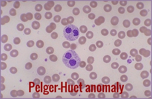
Pelger-Huët Anomaly (PHA) is a rare inherited blood disorder that affects the shape and function of white blood cells (WBCs). Pelger-Huët Anomaly is characterized by abnormal nuclear segmentation of neutrophils and other granulocytes, resulting in a bilobed or peanut-shaped nucleus instead of the normal trilobed shape. Although PHA is usually asymptomatic and benign, it can sometimes be associated with other medical conditions or indicate an underlying genetic disorder.
Pelger-Huët anomaly is caused by a mutation in the LBR (lamin B receptor) gene, which encodes a protein involved in the nuclear envelope structure and function. The mutation leads to abnormal nuclear shape and chromatin organization in WBCs, but not in other cell types. Pelger-Huët Anomaly is inherited in an autosomal dominant pattern, which means that a person with one copy of the mutated gene (heterozygous) has a 50% chance of passing it on to each of their offspring. However, most cases of PHA are sporadic, meaning that the mutation occurs spontaneously in a person with no family history of the disorder.
The characteristic leukocyte appearance was first reported in 1928 by Pelger, a Dutch hematologist, who described leukocytes with dumbbell-shaped bilobed nuclei, a reduced number of nuclear segments, and coarse clumping of the nuclear chromatin.
In 1931 Huet, a Dutch pediatrician, identified it as an inherited disorder.
Neutrophils contain not more than two lobes to the nuclei (spectacle-shaped nuclei or peanut-shaped nuclei instead of the normal trilobed shape) and “band forms” are frequent. In the inherited anomaly, affected neutrophils with bilobed nuclei makeup 60-90% of the neutrophils seen.
PHA can be diagnosed by examining blood smears under a microscope and identifying the characteristic bilobed or peanut-shaped nuclei of neutrophils and other granulocytes. The anomaly can be quantified by calculating the Pelger-Huët ratio, which is the percentage of neutrophils with abnormal nuclei among all neutrophils counted. A ratio of 10% or higher is considered diagnostic of PHA. However, the diagnosis should be confirmed by genetic testing to exclude other causes of nuclear segmentation abnormalities, such as myelodysplastic syndromes (MDS) or acute myeloid leukemia (AML).
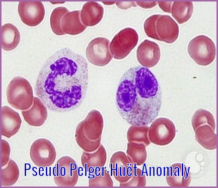
Anomalies resembling Pelger-Huët anomaly that are acquired rather than familial have been described as pseudo Pelger-Huët anomaly (PPHA). The morphologic characteristic seen in pseudo Pelger-Huët anomaly is similar to PHA. The acquired pseudo Pelger-Huët anomaly has been associated with pathologic conditions including acute and chronic myeloid leukemias, myelodysplastic syndrome (MDS), severe infections and some other toxic conditions.

A cluster of pseudo Pelger-Huet neutrophils in the bone marrow aspirate of a patient with myelodysplastic syndrome
Pseudo Pelger-Huët anomaly and micromegakaryocytes are considered to be highly characteristic and highly pathognomonic of MDS.

Blood smear of a patient with myelodysplastic syndrome: red blood cells showing marked poikilocytosis, in part related to post-splenectomy status, a central and hypogranular neutrophil with a pseudo-Pelger-Huet nucleus.
Usually, the congenital form is not associated with thrombocytopenia and leukopenia, so if these features are present more detailed search for myelodysplasia is warranted, as pseudo-Pelger–Huët anomaly can be an early feature of myelodysplasia.
When Pelger-Huët cells are identified, initially attempt to determine if the patient has a benign inherited anomaly or an acquired morphologic feature (ie, pseudo–Pelger-Huët anomaly). In PPHA, cells may appear morphologically similar to inherited PHA, but the absence of these findings in other family members, a low percentage of affected cells (usually 5-20%), and involvement of other cell lines (eg, anemia or thrombocytopenia) suggest an acquired anomaly.
Conclusion:
Pelger-Huët anomaly is usually considered a benign condition that does not require treatment or follow-up, as it does not cause any clinical symptoms. However, PPA can sometimes be associated with other medical conditions or indicate an underlying genetic disorder. For example, PHA has been reported in patients with myeloproliferative neoplasms (MPNs), such as chronic myeloid leukemia (CML) or essential thrombocythemia (ET), as well as in patients with congenital disorders, such as Down syndrome or Alagille syndrome. Therefore, patients with Pelger-Huët anomaly should be evaluated for any signs or symptoms of these conditions and undergo appropriate investigations if necessary.
References:
Peter Maslak and Susan McKenzie. Pseudo Pelger Huet Cells (II) – 1. | ASH Image Bank | American Society of Hematology https://imagebank.hematology.org/image/1848/pseudo-pelger-huet-cells-ii–1 #ASHImageBank
Bain BJ. Blood Cells: A Practical Guide. 5th ed. Wiley-Blackwell; 2015.
Bux J, Behrens G, Jaeger G, et al. Diagnosis and clinical course of autoimmune neutropenia in infancy: analysis of 240 cases. Blood. 1998;91(1):181-186.
Rao LVM, Gowda V. Pelger-Huët anomaly: a rare inherited anomaly of leukocytes. Blood Cells Mol Dis. 2014;53(4):223-227.
Rosinol L, Cibeira MT, Blade J, et al. Pelger-Huët anomaly in multiple myeloma: a new prognostic factor? Eur J Haematol. 2001;66(6):383-386.
Wikipedia contributors. (2022, April 25). Pelger–Huët anomaly. In Wikipedia, The Free Encyclopedia. Retrieved 23:48, November 25, 2023, from https://en.wikipedia.org/w/index.php?title=Pelger%E2%80%93Hu%C3%ABt_anomaly&oldid=1084653058



Mutation here involve which Gene?
Hello,
Thank you for your message.
Pelger-Huët anomaly is a benign, dominantly inherited defect of terminal neutrophil differentiation secondary to mutations in the lamin B receptor (LBR) gene.
Regards,
My brother and my oldest son both have been verified as having PHA. Other things they have in common are Dyslexia and a few other learning issues. Could these possibly be connected somehow? I think I read somewhere that certain mutations in the LBR gene can cause learning issues.
Hi Suzanne,
Thank you for your comment.
I really don’t know if there is a link between PHA and Dyslexia but I think this article may answer some of your queries with this matter:
https://rarediseases.info.nih.gov/diseases/9148/pelger-huet-anomaly
BW,
I went to the hospital recently for heart issue due to over stress. This test came back in my patient portal.
Smear Exam by Pathologist
Note:
Mar 02, 2022 11:18 a.m. EST
Comment:
Red blood cells show minimal variation in shape and size with no inclusions and less than one schistocyte per 100 red blood cells seen. No nucleated red blood cells are seen. The white blood cell differential count is confirmed. Normal morphological features are seen; rare Pelger Huet cells seen. The platelet count shows decreased numbers. No platelet clumping is seen. Peripheral Smear Review: Mild macrocytic anemia NOTE: Vit B12/Folate levels and if normal, considerations for a marrow study is recommended.
Hi Pamela,
Thank you for your comment.
From your blood film comment they are querying ?possible myelodysplasia in view of the macrocytic anaemia and the Pelger-Huët anomaly. I would suggest initially to check your serum B12, Folate, GGT, and LDH.
BW,
Hello, I was wondering if the following results of “rare hypograndular neutrophil” is the same as PHA or APHA.
Normochromic, normocytic RBCs. Platelets adequately granulated. Rare hypogranular neutrophil of uncertain significance in this setting. No other features of dysplasia. No blasts.
Hi Christine,
Thank you for your comment.
As long as the hypogranular neutrophils are not seen frequently on the blood film and your CBC is normal you can safely ignore it.
BW,
Is this “condition “ related to Eosinophilic?
I am not medically educated but fascinated with diagnoses and medical vocabulary.
Thank you for your kind response.
Hi Ila,
Thank you for reaching out.
Pelger-Huët anomaly (PHA) does not directly relate to eosinophilia. PHA is a genetic disorder characterized by abnormal nuclear shape in neutrophils, where the nuclei appear hyposegmented.
This condition is typically benign and does not affect the function of the neutrophils.
Eosinophilia, on the other hand, refers to an elevated number of eosinophils in the blood and can be caused by a variety of conditions, including allergic reactions, parasitic infections, and certain hematologic disorders.
There is no known direct association between PHA and eosinophilia.
Best wishes,
Dr. M. Abdou