PET imaging in Hematology
Positron emission tomography (PET) scans are used to produce detailed 3-dimensional images of the inside of the body. PET imaging in hematology has become a cornerstone procedure which is particularly helpful for investigating confirmed cases of cancer to determine how far cancer has spread and how well it’s responding to treatment.
PET is a nuclear medicine medical imaging technique that produces a 3-D image of functional processes in the body. A PET scan uses a small amount of a radioactive drug, or tracer, to show differences between healthy tissue and diseased tissue. The most commonly used tracer is called FDG (fluorodeoxyglucose), so the test is sometimes called an FDG-PET scan. Before the PET scan, a small amount of FDG is injected into the patient.
Because cancer grows at a faster rate than healthy tissue, cancer cells absorb more of the FDG. The PET scanner detects the radiation given off by the FDG and produces color-coded images of the body that show both normal and cancerous tissue.
A PET scan can show how well certain body parts work rather than simply showing what they look like. The images can clearly show the part of the body being investigated, including any abnormal areas, and can highlight how well certain body functions are working.
PET scans are often combined with CT scans to produce more detailed images. This is known as a PET-CT scan. PET-CT scans are generally considered more accurate in diagnosing cancer than PET scans alone.
With modern dedicated PET machines, a resolution of approximately 5 mm can be achieved. Moreover, PET imaging is more precise than conventional scintigraphy using a quantitative approach to interpretation with the standardized uptake value (SUV) (ratio of activity per volume unit over injected activity per body mass).
An SUV of 2.5 or higher is generally considered to be indicative of malignant tissue; however, there has been a wide range of SUVs reported for similar diseases. It is important to recognize that an SUV around 2.5 can be measured in non-malignant regions. Conversely, small tumors can also exhibit a maximum SUV of < 2.5.
PET-CT scans can help to:
- diagnose cancer.
- stage cancer, so doctors know whether it has spread.
- decide whether you can have surgery to remove your cancer.
- decide which is the best treatment for your cancer.
- find the place in the body where your cancer first started to grow (primary cancer).
- check whether your cancer has come back.
A PET-CT scan can also show how well a cancer treatment is working.
PET scans may also occasionally be combined with an MRI scan (known as a PET-MRI scan).
How PET scans work?
PET scanners work by detecting the radiation given off by a substance injected into the arm called a radiotracer as it collects in different parts of the body.
In most PET scans, a radiotracer called fluorodeoxyglucose (FDG) is used, which is similar to naturally occurring glucose, so the body treats it in a similar way.
By analyzing the areas where the radiotracer does and doesn’t build up, it’s possible to determine how well certain body functions work and identify any abnormalities. For example, a concentration of FDG in the body’s tissues can help identify cancerous cells because cancer cells use glucose at a much faster rate than normal cells. 
The adoption of the PET scan has been most notable in treating lymphomas, where its incorporation into the care of Hodgkin’s and non-Hodgkin’s lymphomas is now virtually universal. PET scans have clearly impacted the way lymphomas are staged and treated, particularly those types with the potential for cure, such as classical Hodgkin’s lymphoma and large cell and/or aggressive lymphoma. 
PET scans, because of their enhanced sensitivity and convenience, have almost totally replaced gallium scans. Unlike gallium scans, PET scans are routinely used in staging since the scans can reveal disease in normal-sized lymph nodes (on CT scan), which would otherwise have been interpreted as unremarkable. 
PET scanning has altered treatment approaches as well. For instance, bulky masses, despite being sterilized of lymphoma, are often characterized by some residual mass or scar. Using conventional means, it has been difficult to determine whether the residual mass after treatment did or did not contain active disease. As a result, these masses had been routinely irradiated even if a biopsy showed a scar (because of possible sampling error). Although many still advocate irradiating bulky masses, the advent of the PET scan has engendered a rethinking of this approach when no disease activity is apparent.
PET Scans in the Staging of Lymphoma:
Staging is important in treating all malignancies but is critically important for patients with lymphoma. Accurate staging allows the minimization of toxic therapies, such as extended-field radiation or overly aggressive chemotherapy, decreasing the risk of secondary malignancies, which exceeds 10% in several historical series of patients with early-stage Hodgkin’s disease. Patient quality of life during and after treatment may also be improved with tailored therapy defined by staging.
Most PET studies involve patients with either Hodgkin’s disease or diffuse large B-cell non-Hodgkin’s lymphoma. PET detects more disease sites above and below the diaphragm on lymphoma staging than gallium scintigraphy and may have particular utility in evaluating the spleen. Perhaps more importantly, PET may help to characterize a residual mass on anatomic imaging following therapy as either fibrosis or residual active lymphoma. Moreover, persistently positive PET scans during and after chemotherapy appear to have a high sensitivity for predicting subsequent relapse. A negative PET scan at the end of therapy provides very favorable prognostic information.
Lymphoma Histology and FDG Uptake:
Most studies evaluating FDG-PET in lymphoma include patients with diffuse large B-cell non-Hodgkin’s lymphoma (NHL) or Hodgkin’s disease. There is limited data available on the role of PET in other histologies. A retrospective review of 172 patients with various types of lymphoma who underwent FDG-PET imaging was completed at the University of Pennsylvania. Only 6% of those patients had no evidence of disease on PET scans. FDG-PET accurately detected disease in patients with diffuse large B-cell NHL, mantle cell lymphoma, follicular lymphoma, and Hodgkin’s disease. PET was less reliable at detecting marginal zone lymphoma, a finding that has been confirmed by other groups, particularly in the case of extranodal marginal zone lymphomas.
In another study, limited to patients with indolent B-cell lymphomas, PET appeared to have the potential to contribute to managing patients with follicular NHL. Still, sites of disease in patients with small lymphocytic lymphomas were only detected approximately 50% of the time. Small series have suggested that using SUV to determine the intensity of FDG uptake, PET may be able to predict the histologic transformation of indolent lymphoma; however, the relationship among pathological subtype, mitotic rate, and SUV remains controversial. 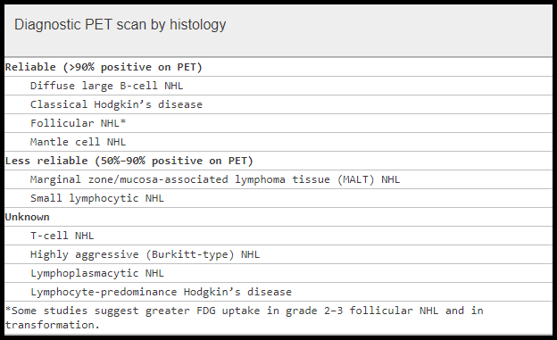
PET CT Scan in Myeloma:
18F-FDG PET/CT is commonly used to detect myeloma bone disease and extra-medullary sites of metabolically active myeloma foci with relatively high sensitivity and specificity. Furthermore, follow-up PET/CT scans can be used to monitor treatment response and are of prognostic value for survival. This imaging modality uses enhanced glucose metabolic activity to visualize areas of interest and detect more lesions than whole-body X-rays. A decrease in 18F-FDG uptake correlates with the chemotherapeutic response, and residual 18F-FDG uptake after completion of therapy may serve as an accurate marker for assessing minimal residual disease (MRD) comparable to next-generation sequencing or multi-colour flow cytometry. According to the International Myeloma Working Group (IMWG) response criteria, developing new bony lesions or soft tissue plasmacytoma on imaging modalities, including PET/CT, indicates progressive disease (PD) independent of serum monoclonal protein or light chain concentration.

The image depicts a myeloma pretreatment PET scan showing widespread axial skeletal disease and diffuse marrow 18F-FDG uptake. Post-treatment PET scan shows the response with therapy in the axial skeleton and bone marrow.
Deauville Criteria:
The Deauville 5-point scoring system is an internationally accepted and utilized five-point scoring system for the FDG avidity of a Hodgkin’s lymphoma or Non-Hodgkin’s lymphoma tumor mass as seen on PET scan:
Score 1: No uptake above the background.
Score 2: Uptake ≤ mediastinum.
Score 3: Uptake > mediastinum but ≤ liver.
Score 4: Uptake moderately increased compared to the liver at any site.
Score 5: Uptake markedly increased compared to the liver at any site.
Score X: New areas of uptake unlikely to be related to lymphoma.
Scores of 1 and 2 are considered to be negative, and 4 and 5 are considered to be positive. “Score 3 should be interpreted according to the clinical context, but in many Hodgkin’s Lymphoma patients, it indicates a good prognosis with standard treatment.”
Preparation:
Your doctor will give you detailed instructions on preparing for your scan. A general rule is to not eat anything for at least 6 hours before the scan. You will be encouraged to drink water. Wear comfortable clothes.
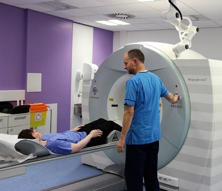
During the Examination:
A nurse or technologist will take you to a special room where you will receive an intravenous (IV) injection of the radioactive drug. Sometimes, you will be asked to inhale the drug instead. Then, you will wait 30 to 90 minutes for the drug to travel through your body and accumulate in the tissues being studied. During this time, you will rest quietly and avoid movement. You won’t be able to feel the drug in your body.
The PET scanner is a large machine with a hole in the middle. It looks like a doughnut with a table in the middle. You will lie on the table. The table will slide into the machine. You will be asked to remain still during the scan.
Time Required:
30 to 45 minutes.
Noise During Examination:
Buzzing or clicking sounds.
Space During Examination:
You will lie on a narrow table that slides into a circular opening of the scanner. The size of the opening is 27 to 30 inches. How much space you feel you have around you will depend on your body size and the scanner used. A mild sedative may be used to help you feel more comfortable during the exam. Let your doctor know if you are anxious about being in enclosed spaces.
PET imaging in hematology
Benefits:
The functional information obtained by a PET scan is unique and unavailable using other types of imaging. For many diseases, PET provides the most useful information required to diagnose and determine the most appropriate treatment.
Risks:
Although a radioactive tracer is used during a PET scan, the amount of radiation you are exposed to is low and short-lived. It is not enough to affect normal body processes. However, there are risks due to the tracer:
- The radioactive substance may expose radiation to the fetus of a pregnant woman or to the infant of a woman who is breastfeeding. If you are pregnant or nursing, please discuss this with your doctor.
- There is a rare risk of a major allergic reaction to the tracer.
Cost of PET Scan:
The cost of a PET scan can vary significantly depending on several factors, including the geographic location, the type of PET scanner used, the complexity of the procedure, and the healthcare provider. Here is a general overview of the cost of PET scans worldwide:
- The average cost of a PET scan ranges from $1,500 to $3,000 in the United States.
- In the United Kingdom, the cost of a PET scan typically falls between £1,000 and £2,000.
- In Canada, the cost of a PET scan is usually covered by provincial health insurance plans.
- In Australia, the cost of a PET scan can range from $1,000 to $2,500.
- In many developing countries, the cost of a PET scan can be significantly higher due to limited access to advanced medical technology and infrastructure.
It’s important to note that these are just approximate ranges, and the actual cost may vary depending on individual circumstances and specific healthcare providers. If you are considering undergoing a PET scan, it is advisable to consult with your healthcare provider or insurance company to obtain accurate information about the cost and coverage options.
References:
Thompson CJ. Instrumentation. In: Wahl RL (Ed.), Principles and Practice of Positron Emission Tomography. Lippincott Williams & Wilkins, Philadelphia, PA, USA; 2002: 48–64.
Coleman M. The Use of PET Scanning in Lymphoma: Progress and Problems. HemOnc Today. September 25, 2009. Available at: https://goo.gl/YFdVhK
Friedberg JW. PET Scans in the Staging of Lymphoma: Current Status. The Oncologist. 2003; 8(5): 438–447. https://theoncologist.onlinelibrary.wiley.com/doi/full/10.1634/theoncologist.8-5-438
Ng AK, Bernardo MV, Weller E, et al. Second malignancy after Hodgkin disease treated with radiation therapy with or without chemotherapy: long-term risks and risk factors. Blood. 2002; 100(6): 1989–1996. doi:10.1182/blood.V100.6.1989
Elstrom R, Guan L, Baker G, et al. Utility of FDG-PET scanning in lymphoma by WHO classification. Blood. 2003; 101(10): 3875–3876. doi:10.1182/blood-2002-11-3546
Hoffmann M, Kletter K, Diemling M, et al. Positron emission tomography with fluorine-18-2-fluoro-2-deoxy-D-glucose does not visualize extranodal B-cell lymphoma of MALT type. Ann Oncol. 1999; 10(10): 1185–1189. doi:10.1023/A:1008394900859
Jerusalem G, Beguin Y, Najjar F, et al. PET with 18F-FDG for the staging of low-grade non-Hodgkin’s lymphoma. Ann Oncol. 2001; 12(6): 825–830. doi:10.1023/A:1011197427507
Barrington SF, et al. Role of imaging in the staging and response assessment of lymphoma: Consensus of the International Conference on Malignant Lymphomas Imaging Working Group. Eur J Nucl Med Mol Imaging. 2010; 37(9): 1824–1833. doi:10.1007/s00259-010-1428-0
American College of Radiology Imaging Network (ACRIN). About PET Scans. https://www.acrin.org/PATIENTS/ABOUTIMAGINGEXAMSANDAGENTS/ABOUTPETSCANS.aspx
Follows GA, et al. Guidelines for the first-line management of classical Hodgkin lymphoma. Br J Haematol. 2014; 166(1): 34–49. doi:10.1111/bjh.12878
Mah K, Caldwell CB. PET-CT in Radiotherapy Treatment Planning. In: Standardized Uptake Value – ScienceDirect Topic Overview. Elsevier; 2008. https://www.sciencedirect.com/topics/medicine-and-dentistry/standardized-uptake-value
University of Edinburgh. What Is a PET-CT Scan? https://www.ed.ac.uk/clinical-sciences/edinburgh-imaging/for-patients-study-participants/tell-me-more-about-my-scan/what-is-a-pet-ct-scan
Sundaram S, Driscoll J, Fernandez-Ulloa M, de Lima M, Malek E. FDG PET imaging in multiple myeloma: implications for response assessments in clinical trials. Am J Nucl Med Mol Imaging. 2018; 8(6): 421–427. PMID: 30697462; PMCID: PMC6334211
Durie BG, Waxman AD, D’Agnolo A, Williams CM. Whole-body 18F-FDG PET identifies high-risk myeloma. J Nucl Med. 2002; 43(11): 1457–1463. https://jnm.snmjournals.org/content/43/11/1457
Bartel TB, Haessler J, Brown TL, et al. F18-FDG PET in context of other imaging and prognostic factors in multiple myeloma. Blood. 2009; 114(10): 2068–2076. doi:10.1182/blood-2009-03-213280
Kumar S, Paiva B, Anderson KC, et al. International Myeloma Working Group consensus criteria for response and minimal residual disease assessment in multiple myeloma. Lancet Oncol. 2016; 17(8): e328–e346. doi:10.1016/S1470-2045(16)30206-6
Keywords:
PET CT scan, PET imaging in hematology, FDG PET, PET scan for cancer, PET scan meaning, difference between PET scan and CT scan, PET CT meaning, PET scan for cancer patients, PET scan cost, whole-body PET, standardized uptake value SUV, FDG uptake, lymphoma staging, Deauville score, Hodgkin lymphoma, non-Hodgkin lymphoma NHL, multiple myeloma imaging, PET scan in myeloma, PET CT in hematology, PET scan for cancer diagnosis, minimal residual disease MRD myeloma, therapeutic response monitoring, PET MRI scan, what is a PET CT scan, what is a PET scan used for, what is a PET scan used to diagnose, what is a PET scan like, how PET CT is done, difference between PET CT and CT, PET and CT difference, PET imaging meaning, PET CT scan in myeloma diagnosis, PET imaging cost, PET CT scan cost, FDG PET in lymphoma, FDG PET in multiple myeloma, PET CT for radiotherapy planning, PET imaging in blood cancer, functional imaging hematology, PET CT interpretation guidelines, PET scan vs MRI, PET CT results explained

Related Posts:


 Cells")
Request Online Consultation With Dr M Abdou
Fee: US$100
Secure payment via PayPal (credit and debit cards accepted)
Pay Now

I appreciate you helping me learn more how a PET scan works. It is interesting that it can detect the radiation given off by the injected liquid in the body. My nephew is thinking of becoming a scanning technician. He might interested in knowing this.
Hi Elisabeth,
Thank you for your comment.
The PET scan uses a special dye containing radioactive tracers. These tracers are either swallowed, inhaled, or injected into a vein in your arm depending on what part of the body is being examined. Certain organs and tissues then absorb the tracer. When detected by a PET scanner, the tracers help your doctor to see how well your organs and tissues are working. The tracer will collect in areas of higher chemical activity, which is helpful because certain tissues of the body, and certain diseases, have a higher level of chemical activity. These areas of disease will show up as bright spots on the PET scan. The PET scan can measure blood flow, oxygen use, how your body uses sugar, and much more.
Regards,
Dr. Abdou,
My mother is 96 and many test have been done to figure out what is wrong with her lung. Everything has been ruled out except cancer, but after 3 Cscans they can’t make a diagnosis. Would you reccomend a PET scan? They are talking about taking fluid from the outside of her lung to test it for cancer cells. She doesn’t want the procedure where they take specimens from inside the lungs. The pulmonary doctor seems to think because she is 96 just let it fo and die. Would like to hear what you think. Thank you.
Nancy
Hi Nancy,
Thank you for your message.
The PET scan uses a mildly radioactive drug to show up areas of the body where cells are more active than normal. It’s used to help diagnose some conditions including cancer. It can also help to find out where and whether cancer has spread. I encourage doing a PET-CT scan and a CT guided biopsy for your mother to reach a definite diagnosis if she is a good 96 i.e. without multiple comorbidities and has a reasonable performance status (0-2) as long as she can go for the test as some cancers may respond well to treatment like lymphomas for example.
Best wishes,
I have a lot of small dots of blood and medium and large bruises for no reason when I do hit myself instantly I bruise
I am very tired no energy.. no blood thinners. Primary M.D. just tells me I have low red blood cells. Should I request to see a hematologist
?
Hi Caroline,
Thank you for your message.
I would suggest initially to check your FBC, Coagulation Screen, Serum iron/ferritin, B12, and Folate.
Best wishes,
Good evening doctor
My 4 years old son has been diagnosed with NHL (Burkitt) with multiple hepatic liver masses but his bone marrow is negetive now he is reciveing the third cycle (mtx , cytosar) , he completed 2 induction cycle(cyclophosmide , mtx , adrimycin ,vencstrin and rituxmab )and i have some question
1-does liver involved in burkitt lymphoma?
2-is it stage 3 or 4 ?
3-how is the prognoses of burkitt lymphoma with this state ?
FYI he is feeling better now
with recpect
Hi Adnan,
Thank you for your message and wishing your son a speedy recovery Insha Allah.
1- Any organ can get involved.
2- I’ll classify it as stage 4 even if the bone marrow is spared.
3- Prognosis is good and he may achieve a complete cure with treatment.
BW,